Isoelectric Focusing Electrophoresis Service
- Biopharmaceutical Research and Development
- Clinical Diagnostics
- Food and Beverage Quality Control
- Environmental Monitoring
- Academic and Basic Research
- Biotechnology Product Characterization
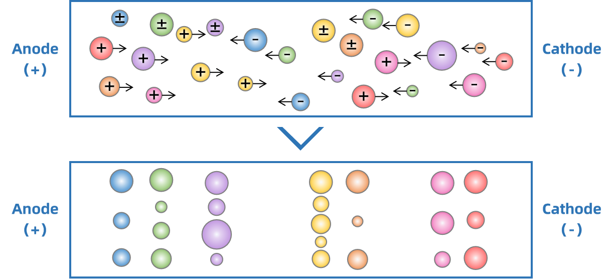
Isoelectric focusing electrophoresis (IEF) is a highly precise analytical technique used to separate proteins based on their isoelectric point (pI)—the pH at which a protein carries no net electrical charge. In an electric field applied across a stable pH gradient, proteins migrate based on their net charge. Positively charged proteins move towards the negatively charged cathode, while negatively charged proteins move towards the positively charged anode. As a protein travels through the pH gradient, it encounters a region where the surrounding pH matches its isoelectric point (pI)—the pH at which its net charge becomes zero. At this point, the protein no longer experiences an electrical driving force because the attractive and repulsive electrostatic forces acting on it are perfectly balanced, and as a result, the protein stops migrating. This unique principle allows isoelectric focusing electrophoresis to achieve exceptionally high resolution, effectively separating proteins with even minor differences in their pI values. Techniques such as slab gel IEF, capillary IEF, and immobilized pH gradient (IPG) gels further refine this separation, ensuring accuracy and reproducibility in protein charge profiling.

Figure 1. The Principle of Isoelectric Focusing Electrophoresis
IEF plays a crucial role in biopharmaceutical research and manufacturing. Isoelectric focusing electrophoresis is widely applied in protein heterogeneity analysis, charge variant profiling, quality control of therapeutic proteins, and identification of post-translational modifications such as deamidation, phosphorylation, and glycosylation. Additionally, isoelectric focusing electrophoresis is routinely employed in stability studies, formulation optimization, and batch-to-batch consistency evaluation. Its compatibility with downstream analytical techniques, such as mass spectrometry (MS) and immunoassays, further extends its utility, enabling a more comprehensive understanding of protein structure and function. These capabilities make IEF an essential component in regulatory submissions, ensuring that therapeutic proteins meet stringent safety and efficacy standards.
Service at MtoZ Biolabs
MtoZ Biolabs offers a comprehensive Isoelectric Focusing Electrophoresis Service that provides high-resolution separation and characterization of proteins based on their isoelectric points, enabling detailed analysis of charge variants, protein purity, and post-translational modifications. Our service covers every stage of the analytical workflow, including experimental design, precise sample preparation, high-sensitivity IEF separation, and thorough data interpretation. By leveraging cutting-edge platforms, we ensure accurate identification of protein heterogeneity, structural integrity, and functional stability. Whether you are conducting early-stage characterization, optimizing biopharmaceutical formulations, or preparing regulatory submissions, MtoZ Biolabs’ Isoelectric Focusing Electrophoresis Service delivers reliable, reproducible, and actionable results.
Analysis Workflow
1. Sample Preparation
In the initial step, proteins are extracted, purified, and adjusted to optimal concentrations to meet isoelectric focusing electrophoresis analysis requirements.
2. pH Gradient Selection and Optimization
The appropriate pH gradient is selected based on the protein's isoelectric point (pI) range and biochemical characteristics. Immobilized pH gradient (IPG) gels or ampholytes are calibrated to ensure stability and reproducibility throughout the isoelectric focusing electrophoresis process. Gradient ranges are optimized to maximize separation resolution and prevent overlapping protein bands.
3. Isoelectric Focusing (IEF) Separation
An electric field is carefully applied across the pH gradient, guiding proteins to migrate until they reach their isoelectric points (pI). To ensure optimal separation, key parameters such as voltage, current, and temperature are tightly regulated, minimizing the risk of protein diffusion or degradation. Throughout the process, real-time monitoring guarantees precise protein focusing, preventing over-migration or band smearing and ensuring reliable, high-resolution results.
4. Visualization and Analysis
After separation, proteins are visualized using staining techniques such as Coomassie Blue or silver staining, or via fluorescent labeling for enhanced sensitivity. High-resolution imaging systems capture clear and detailed electropherograms, ensuring accurate representation of protein separation patterns. In the analysis phase, charge variant distribution, protein heterogeneity, and key post-translational modifications are assessed, providing a reliable foundation for further data interpretation.
Service Advantages
1. Advanced Analysis Platform: MtoZ Biolabs established an advanced Isoelectric Focusing Electrophoresis Service platform, guaranteeing reliable, fast, and highly accurate analysis service.
2. One-Time-Charge: Our pricing is transparent, no hidden fees or additional costs.
3. High-Data-Quality: Deep data coverage with strict data quality control. AI-powered bioinformatics platform integrates all Isoelectric Focusing Electrophoresis Service data, providing clients with a comprehensive data report.
4. Customized Workflow Design: Each project is tailored to meet specific client needs, from sample preparation to reporting, ensuring alignment with diverse research, development, and quality control goals.
Applications
Isoelectric focusing electrophoresis is widely applicable across various fields, offering precise protein characterization analysis.
Case Study
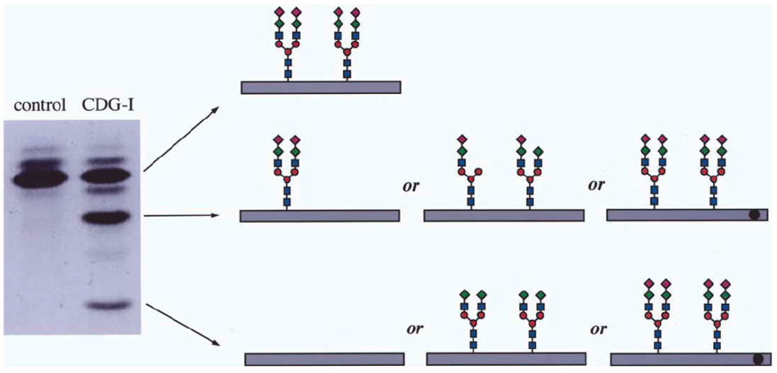
Isoelectric focusing electrophoresis of serum transferrin is a key screening tool for congenital disorders of glycosylation (CDG). In healthy controls, the dominant band represents tetrasialotransferrin, while CDG-I patients show additional bands corresponding to disialo- and asialotransferrin due to incomplete glycosylation. In this case, isoelectric focusing electrophoresis successfully detected abnormal migration patterns in a CDG-I patient, indicating disrupted glycan structures. While IEF cannot determine the exact glycan alterations, Isoelectric focusing electrophoresis remains a highly sensitive diagnostic tool especially when combined with downstream techniques like mass spectrometry for comprehensive analysis.

Figure 2. Isoelectric Focusing Electrophoresis of Serum Transferrin
FAQ
Q: How do you ensure the stability of the pH gradient and the consistency of the electrophoresis process during Isoelectric Focusing Electrophoresis Service?
MtoZ Biolabs ensures the stability of the pH gradient and the consistency of the electrophoresis process through a combination of advanced instrumentation, optimized protocols, and rigorous quality controls. We utilize high-precision pH gradient generation systems, such as immobilized pH gradient (IPG) gels and carefully calibrated buffers, to maintain a stable and reproducible pH environment throughout the entire analysis. Furthermore, our equipment is regularly calibrated and monitored for voltage stability, temperature control, and uniform electric field distribution to prevent deviations during protein migration. Each experiment includes system suitability tests and standard reference proteins to validate the stability and reproducibility of the pH gradient. These measures collectively ensure that our Isoelectric Focusing Electrophoresis Service delivers high-resolution, reliable, and repeatable results, meeting both scientific and regulatory standards.
How to order?


