



Label-free quantification (LFQ) estimates relative protein abundance from mass spectrometry signals without adding isotopic or chemical labels during sample preparation. Each digest is acquired as its own liquid chromatography-tandem mass spectrometry (LC-MS/MS) run. Software then matches the same peptides across runs, extracts chromatographic peak areas or related intensity features, corrects technical scale differences, and summarizes peptide evidence into protein-level values. The valid comparison is the same protein across samples that share one coherent acquisition and processing plan. LFQ intensities are not molar concentrations and cannot rank different proteins by copy number. The route is useful when labeling chemistry, multiplex channel count, or collection timing would constrain the cohort, and when chromatography can stay stable enough for cross-run alignment.
Researchers who already have defined samples, comparison groups, or a specific project objective can also review the MtoZ Biolabs Label-Free Quantitative Proteomics Service for project-specific feasibility and analysis planning.
When Label-Free Quantification Fits a Study Design
LFQ is a relative discovery strategy for independently injected samples. The first decision is whether the scientific endpoint needs that kind of comparison, not whether an unlabeled workflow sounds simpler than tandem mass tags (TMT) or stable isotope labeling by amino acids in cell culture (SILAC).
Research questions LFQ can support
LFQ can support treatment, genotype, time-point, or tissue-state contrasts in which the same protein is compared across groups. After alignment, normalization, and statistical review, a higher protein-level value can support a relative abundance change. It cannot report copies per cell, a calibrated tissue concentration, or a diagnostic cutoff.
A predefined peptide panel with a calibration curve belongs to a targeted assay. Many conditions that must share one pooled injection belong to isobaric labeling. LFQ covers the remaining need: broad relative discovery, one sample per run, no labeling chemistry.
Sample and cohort conditions that favor unlabeled analysis
Unlabeled analysis is often workable when samples cannot enter a labeling reaction cleanly, arrive over a planned acquisition window, or exceed a fixed multiplex set. Clinical tissues, biofluids, and archival material are common cases: metabolic labeling is unavailable, and chemical labeling may consume material or fail in difficult buffers.
Independence has a cost. Every sample carries its own loading, chromatography, and instrument-response history. Group definitions, injection order, and the intended contrast should be fixed before the first sequence is queued. Identification still requires a searchable protein database or an approved custom database.
How Label-Free Quantification Turns MS Signal into Protein Values
The working assumption is narrow. Under comparable LC-MS conditions, the recorded signal of a given peptide tracks the relative amount of that same peptide. The assumption does not say that two different peptides with similar intensity represent similar protein copy numbers, because digestion recovery, chromatography, ionization efficiency, and interference differ by sequence.
From peptide ion intensity to a protein-level matrix
Proteins are extracted and digested, usually with trypsin, into peptides. Liquid chromatography separates the mixture. The mass spectrometer records precursor ions and fragment spectra. Peptide identities come from fragment evidence searched against a protein database. Quantitative values, in current discovery LFQ, usually come from the precursor chromatogram of an identified peptide: software reconstructs the elution profile and integrates intensity across that window (Cox et al. 2014).
A larger peak area means more precursor signal was recorded for that feature in that run, subject to loading, ionization, interference, and peak shape. Protein values are inferred from peptide evidence. Shared peptides, peptide filtering, missing observations, and the summarization rule can all change the reported protein number.
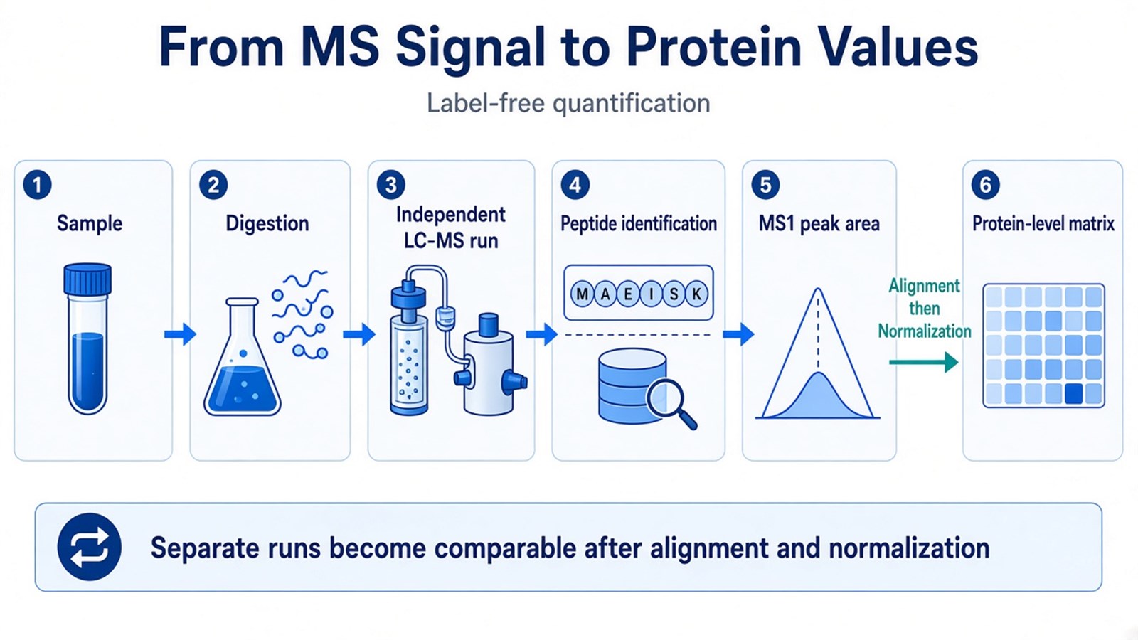
Figure 1. Unlabeled samples stay as separate LC-MS runs; peptide identities and MS1 peak areas become comparable protein values only after alignment and normalization.
Why independently acquired runs need alignment and normalization
Because samples are not chemically coded into one pooled mixture, comparability is created after acquisition. The same peptide can elute at slightly different retention times in different injections. Alignment models those shifts so that features with compatible mass and aligned retention time can be compared. Stable chromatography supports credible correspondence. Column aging, changing peak shape, or altered mobile phases make that correspondence harder to defend.
Normalization addresses systematic differences such as sample loading or overall analytical response. Without it, a globally stronger run can make many proteins appear increased. Building on intensity-based label-free analysis (Cox and Mann 2008), MaxLFQ uses delayed normalization and peptide-ratio information to estimate protein profiles from observed peptide relationships (Cox et al. 2014). Normalization does not repair a confounded run order or stitch datasets acquired on different columns or instruments.
LFQ compares the same protein across independently acquired runs after alignment and normalization.
Label-Free Quantification Methods and How to Choose
Peak area, spectral counting, data-independent acquisition (DIA), and multiple reaction monitoring (MRM) are often listed as if they were interchangeable LFQ recipes. They are not. Labeling, acquisition, and the quantitative metric are separate decisions. Unlabeled sample preparation can be paired with data-dependent acquisition (DDA) or DIA. DDA and DIA name how the instrument schedules fragmentation, not whether a label was used.
Decision layer |
Common options |
What the researcher is choosing |
Do not treat as |
|---|---|---|---|
Labeling |
Unlabeled vs SILAC, TMT, or other tags |
Whether samples stay separate or are coded and pooled |
An acquisition mode |
Quantitative metric |
MS1 peak area; spectral counting; DIA fragment chromatograms |
Which signal becomes the abundance value |
A complete study design |
Acquisition |
DDA or DIA |
How precursors are fragmented and recorded |
Proof that the experiment is unlabeled |
Assay scope |
Discovery vs MRM or PRM |
Proteome-wide relative change vs predefined targets |
Another discovery LFQ algorithm |
Ion-current or total-ion-current comparison is a form of intensity-based quantification, not a third LFQ family.
Intensity-based MS1 peak-area quantification
For DDA-based unlabeled discovery, MS1 peak area is the primary metric. After identification, the precursor extracted-ion chromatogram is integrated and compared across aligned runs. The method supports relative change of the same peptide, then the same protein, across the cohort, and remains sensitive to missing identifications, overlapping features, and chromatographic drift. In MtoZ unlabeled discovery offerings, DDA-LFQ is typically reported from MS1 peak-area workflows rather than from spectral counts.
Spectral counting
Spectral counting estimates protein abundance from the number of assigned fragment spectra. It can help with rough ranking or reanalysis of identification-centric DDA files. Precision is limited when few spectra support a protein, and count-based ratios compress real differences for highly observed proteins. Protein-level fold changes for downstream statistics should use peak-area LFQ.
Unlabeled DDA versus unlabeled DIA
DDA selects precursors in real time for fragmentation. Stochastic sampling is a major source of missing values in unlabeled DDA matrices, especially at low abundance. DIA fragments precursors in predetermined windows and extracts fragment-ion chromatograms, which can improve completeness in larger unlabeled cohorts (Gillet et al. 2012). An unlabeled digest can be run as DIA; a labeled digest can also be acquired with DIA. Acquisition and labeling remain separate decisions.
Unlabeled DIA has its own library, analysis, and missing-value behavior.
Researchers comparing acquisition routes can refer to Label-Free DIA Quantitative Proteomics for library use, analysis, and missing-value behavior in unlabeled DIA.
Targeted unlabeled assays are a separate decision
MRM and parallel reaction monitoring (PRM) quantify predefined precursor-product transitions. They can be performed without isotopic labels, but they ask whether selected peptides change as expected, not whether the proteome-wide matrix is complete. They should not be listed as a fifth LFQ algorithm beside peak area.
How to Read Label-Free Quantification Results
An LFQ table is a matrix of relative values, not a concentration certificate. Interpretation starts from the comparison that the design can actually support.
What an LFQ intensity can support
After alignment, normalization, and protein summarization, an LFQ intensity can support a relative statement: protein A is higher or lower in group 1 than in group 2 within this study. Peptide support, replicate agreement, missingness, and the stated normalization belong next to that statement. A large fold change on one poorly shared peptide is weaker evidence than a smaller change supported by several unique peptides with complete observations.
Ordinary LFQ intensities should not rank protein A against protein B by abundance, infer molar stoichiometry, or transfer a ratio from one instrument or processing pipeline to another study as a calibrated quantity.
An LFQ intensity supports relative change of one protein inside one study, not a copy-number ranking across proteins.
Missing values, batch structure, and run order
Missing values are expected in unlabeled DDA. A low-intensity peptide may be identified in one run and not another. Alignment and Match Between Runs can close some gaps; they do not prove that a protein is absent. Imputation inserts a model-based value, so the method and its effect on group differences should be disclosed.
Injection order matters because chromatographic and instrument performance can change over a sequence. Randomizing biological groups across run order keeps condition and time from becoming the same variable. Quality-control injections can monitor drift, but they cannot rescue a design in which all controls were acquired before all treated samples.
When Another Quantitative Strategy Is the Better Fit
Reassess unlabeled discovery when the endpoint requires a calibrated amount, a predefined-target assay, or near-complete values for low-abundance proteins, or when the cohort cannot stay under one chromatographic plan.
Isobaric labeling, such as TMT, trades labeling chemistry and channel structure for same-run comparison. SILAC is limited to systems that incorporate heavy amino acids. Targeted MS follows a short candidate list; it is not a competing discovery scan.
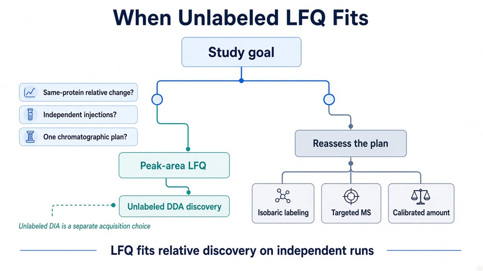
Figure 2. Peak-area LFQ fits relative discovery on independently injected samples; pooled multiplex comparison or predefined targets point to a different method.
When sample number, multiplexing, or acquisition strategy makes the choice less straightforward, researchers can refer to Comparing LFQ and Isobaric Labeling Strategies in Proteomics Quantification for a more detailed strategy-selection framework.
Frequently Asked Questions
1. Is label-free quantification the same as DIA?
No. LFQ means isotopic or isobaric labels were not used. DIA means the mass spectrometer fragments precursors in scheduled windows. Unlabeled samples are often acquired as DIA in discovery work, but DIA is not defined by the absence of a label.
2. Should a study use peak-area LFQ or spectral counting?
Peak-area LFQ is the default for relative discovery statistics. Spectral counting can still help with rough abundance ranking or older DDA identification files. If protein-level fold changes will drive the biological claim, use peak-area values and report peptide support and missingness with them.
3. Can LFQ report absolute protein concentration?
Not from ordinary LFQ intensities. Absolute or copy-number claims need spike-in standards, a calibration model, or a targeted quantitative assay. LFQ supports relative change of the same protein across comparable runs.
4. Can additional samples be acquired after the first LFQ block?
Additional unlabeled samples can be injected when the analytical regime remains compatible. Column replacement, instrument change, or a long interruption can create a new block that ordinary normalization cannot merge into the original cohort. If later enrollment is expected, include that plan before the first acquisition sequence.
Once the comparison design, unlabeled sample set, and expected protein-level readout are defined, the project can be evaluated as an LFQ study. Researchers can review the MtoZ Biolabs Label-Free Quantitative Proteomics Service for project-specific sample evaluation, feasibility assessment, and workflow planning.
