Resources
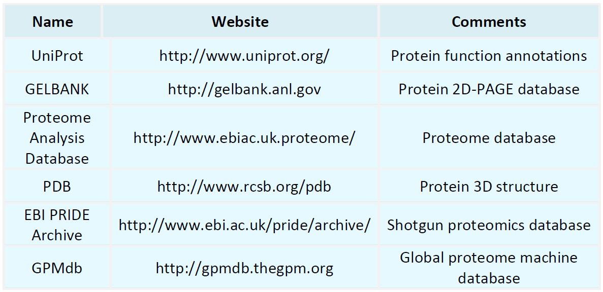
Proteomics Databases
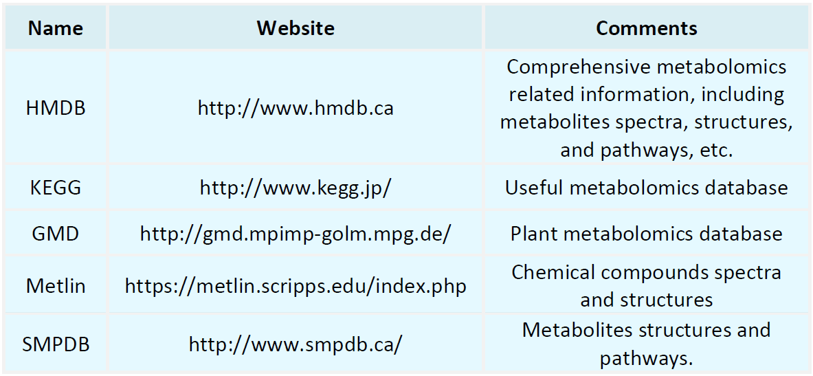
Metabolomics Databases
-
• Classification of Circular Dichroism Spectra
Near-UV Circular Dichroism (Near-UV CD): Near-UV CD measures spectra in the range of 250-300 nm and is mainly used to analyze the tertiary structure of proteins and interactions between subunits. In this wavelength range, the side chains of aromatic amino acids and disulfide bonds have significant absorption peaks, which can be used to study the conformational changes and interactions of proteins.
-
• Circular Dichroism Analyzes Protein Secondary Structure Content
In the field of biochemistry, the assessment of protein secondary structure content represents a fundamental aspect of understanding protein function and stability. The secondary structure of proteins encompasses local conformations, primarily including α-helices, β-sheets, β-turns, and random coils. The unique combinations and spatial arrangements of these structures are critical determinants of the overall three-dimensional architecture and functional capabilities of proteins.
-
• Can Phosphorylation Mass Spectrometry Detect Phosphorylation Sites
Mass spectrometry-based phosphorylation analysis is a powerful technique for identifying phosphorylation sites and quantifying protein phosphorylation. Phosphorylation represents a critical post-translational modification that plays a central role in regulating numerous biological processes, such as the cell cycle, signal transduction, cell differentiation, and metabolism.
-
• Detection of the Isoelectric Point of Glycoproteins
Glycoproteins are ubiquitous in living organisms and are complex macromolecules composed of proteins and carbohydrate moieties. The isoelectric point (pI) of a glycoprotein is the pH at which the molecule is electrically neutral in solution. This pI is typically determined through isoelectric focusing (IEF), an electrophoretic technique performed along a pH gradient. Proteins migrate through this gradient and cease movement upon reaching their pI due to the neutralization of their net charge, allowing for..
-
• Considerations for Detecting Phosphorylated Proteins Using Western Blot
Protein Phosphorylation Detection using Western Blot (WB) is a widely utilized technique in biological research for evaluating protein levels and modifications. Key considerations during the detection process include sample processing, the selection of specific antibodies, the choice of control samples, signal detection, and result interpretation.
-
• Protein Post-Translational Modification Mass Spectrometry Results Analysis
Post-Translational Modifications (PTMs) of proteins occur after translation, involving enzymatic addition or removal of chemical groups such as phosphate, acetyl groups, uridylic acid, and glycosyl groups. These modifications significantly influence protein stability, activity, and functionality. Mass spectrometry serves as a fundamental tool for the analysis of protein PTMs, and the analysis of mass spectrometry data comprises several steps:
-
• How Many Cells Are Needed for Glycosylation Mass Spectrometry Analysis
The number of cells required for glycosylation mass spectrometry analysis is contingent on cell type and the sensitivity of the analysis technique. Past studies typically necessitate millions to billions of cells to obtain a sufficient quantity of glycosylated proteins. The procedures for this analysis encompass cell culture, cryopreservation, cell lysis, and protein extraction. Researchers are advised to thoroughly review pertinent experimental protocols relative to their specific experimental objectives..
-
• Are Post-Translational Modifications of Proteins Reversible
Reversible posttranslational modifications (PTMs) of proteins, such as phosphorylation, acetylation, and ubiquitination, are regulated by specific enzymes that add or remove these chemical groups during cellular physiological processes, thereby ensuring their reversibility. In contrast, irreversible modifications, including glycosylation, lipidation, and methylation, typically alter protein structure and function permanently, as they cannot be removed once established.
-
• Detection of Protein Oxidation Stress Modifications
Protein oxidative stress modifications represent a critical mechanism by which biological systems respond to various biological and environmental stresses. Assessing these modifications provides insight into the organism's health and disease progression, facilitating understanding of the adaptive mechanisms to environmental changes and aiding in the development and optimization of disease prevention and treatment strategies.
-
• Lowry Method for Protein Content Measurement
The Lowry method is a widely adopted technique for quantifying protein concentration, initially introduced by Lowry et al. in 1951. The method is premised on the reaction of proteins with copper ions in an alkaline medium, followed by an oxidation-reduction reaction with the Folin-Ciocalteu reagent, resulting in a color change. The intensity of the resultant color is directly proportional to the protein concentration and can be quantified using a spectrophotometer at a wavelength of 750 nm.
How to order?