


Resources
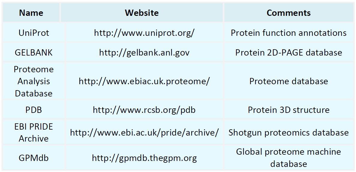
Proteomics Databases
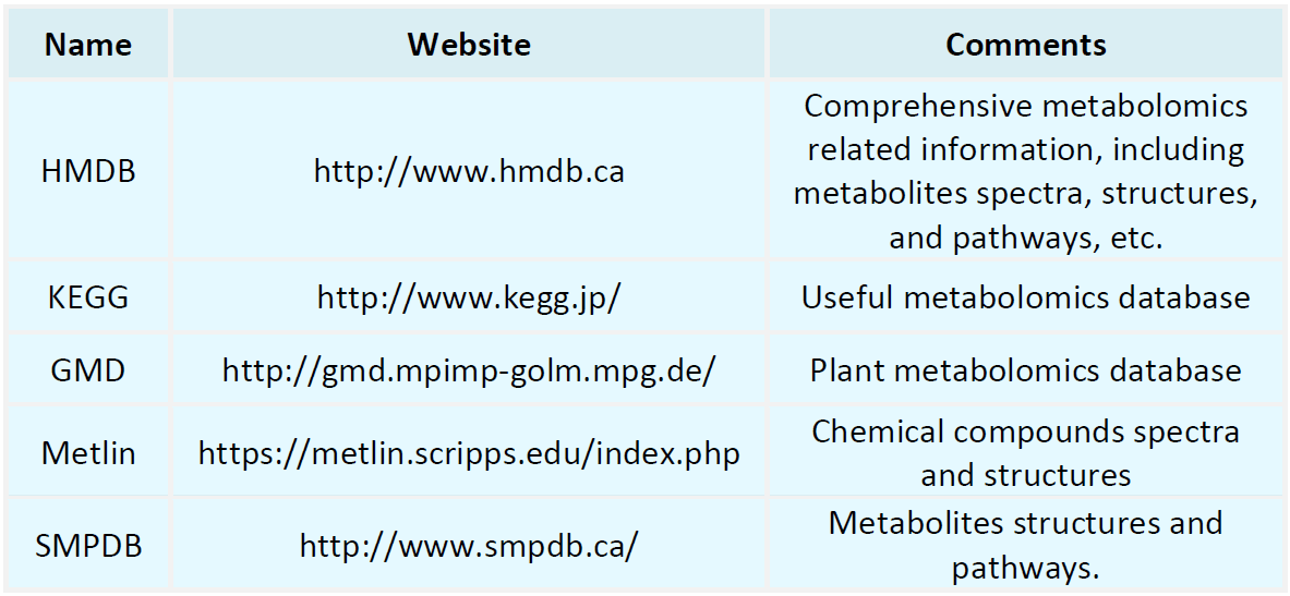
Metabolomics Databases
-
The Edman sequencing method is a classical biochemical technique developed by Pehr Edman in the 1950s for determining the amino acid sequence of proteins. This method employs a stepwise chemical process to sequentially cleave amino acids from the N-terminus of a polypeptide chain. Each amino acid is subsequently separated and identified using chromatographic techniques, enabling the reconstruction of the protein's sequence. The core strength of the Edman sequencing method lies in its precise step-by-s......
-
• LC-MS/MS Protein Quantification
LC-MS/MS protein quantification combines the high-resolution separation capabilities of liquid chromatography (LC) with the sensitivity and precision of tandem mass spectrometry (MS/MS). This technique enables the comprehensive separation, identification, and quantification of complex protein mixtures, providing insights into protein expression levels, post-translational modifications, and interactions in biological systems. Widely used across biomedical research, drug development, disease diagnosti......
-
LC-MS/MS peptide sequencing is a powerful analytical method for identifying and analyzing peptide sequences in biological samples. By combining liquid chromatography (LC) for peptide separation with tandem mass spectrometry (MS/MS) for detailed sequence analysis, this technique provides a robust platform for studying complex proteomes. In LC, peptides are separated based on their retention times, while MS/MS generates peptide fragmentation spectra that allow the determination of their amino acid seque......
-
• Mass Spec Peptide Sequencing
Mass spec peptide sequencing is a highly sensitive and precise technique for analyzing proteins by sequencing their peptides to determine amino acid sequences. This method, a cornerstone of proteomics research, is particularly valuable for identifying and characterizing protein components in complex biological samples. It works by enzymatically or chemically breaking down proteins into smaller peptides, which are then analyzed for their mass using advanced mass spectrometry instruments. Widely regar......
-
• Mass Spectrometry Antibody Characterization
Mass spectrometry antibody characterization is a powerful analytical method used to investigate the structure, composition, and modifications of antibodies at the molecular level. Antibodies, as essential components of the immune system, possess complex structures and diverse functions. This technique provides detailed insights into amino acid sequences, glycosylation patterns, and other post-translational modifications (PTMs), enabling a deeper understanding of antibody functionality and mechanisms. ......
-
• Mass Spectrometry Amino Acid Sequencing
Mass spectrometry amino acid sequencing is a highly sensitive and precise analytical method for determining the amino acid sequences of proteins and peptides. By leveraging the high resolution and sensitivity of mass spectrometry, this technique enables the detailed analysis of complex biological samples to elucidate protein structures. Amino acid sequences are the fundamental components of proteins, and understanding their arrangement is crucial for exploring protein functions, structures, and intera......
-
• Peptide Sequencing Tandem Mass Spectrometry
Peptide sequencing tandem mass spectrometry is a highly sensitive and high-throughput analytical technique used to determine the amino acid sequences of proteins and peptides. By combining multi-stage mass analysis with specific peptide fragmentation patterns, this method enables the resolution of sequence information in complex protein samples. Unlike traditional Edman degradation, peptide sequencing tandem mass spectrometry can directly analyze complex mixtures, making it particularly suitable for s......
-
• Peptide Sequencing by Edman Degradation
Peptide sequencing by Edman degradation is a well-established and highly effective method for determining the amino acid sequences of peptides and proteins. First introduced by Pehr Edman in 1950, this technique employs a stepwise chemical process to selectively cleave and identify N-terminal amino acids in a peptide chain. Through repeated cycles of cleavage and detection, Edman degradation enables precise determination of the sequential arrangement of amino acids, making it an invaluable tool in pro......
-
• Protein Analysis by LC-MS/MS
Protein analysis by LC-MS/MS is a cornerstone of modern proteomics, leveraging the high-resolution separation capability of liquid chromatography (LC) and the sensitivity and precision of tandem mass spectrometry (MS/MS). This technique enables high-throughput identification and quantification of proteins in complex biological samples. It allows the characterization of thousands of proteins and their post-translational modifications (PTMs), offering valuable insights into dynamic changes and regulator......
-
Urine proteomics focuses on the comprehensive analysis of proteins in urine samples using advanced high-throughput technologies. As a non-invasive and easily accessible biological fluid, urine serves as a valuable source for identifying biomarkers linked to physiological and pathological conditions. The primary objective of urine proteomics is to identify and quantify urinary proteins and their modifications, offering insights into their variations under different health states. This approach supports......
How to order?

