


Resources
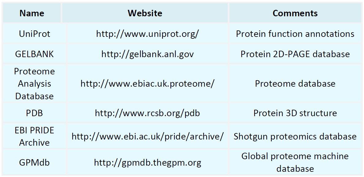
Proteomics Databases
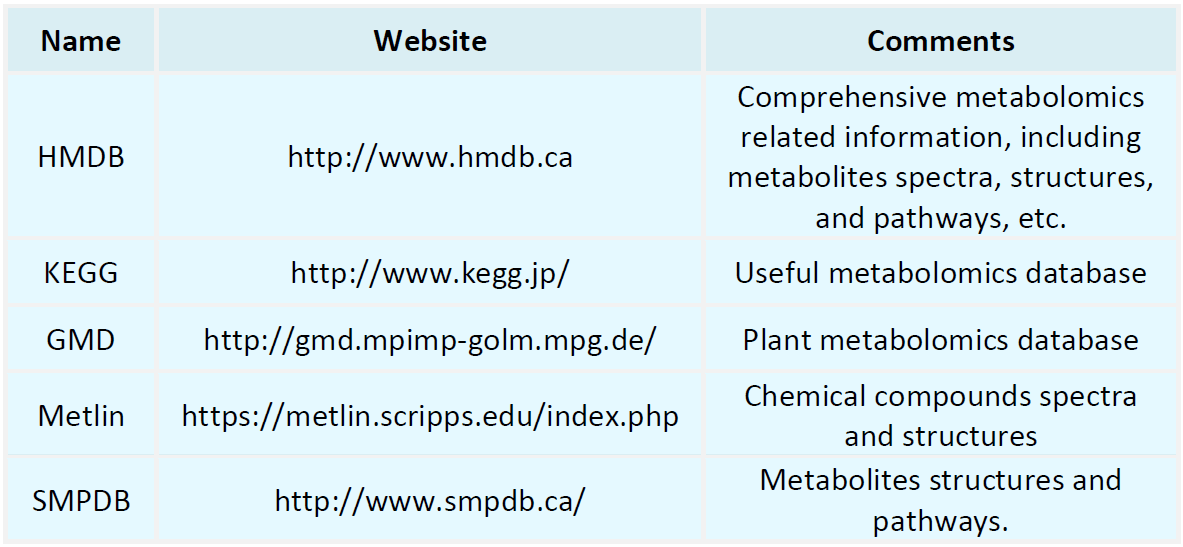
Metabolomics Databases
-
• Subcellular Proteomics Workflow (Including Mass Spectrometry and SILAC Approaches)
In the post-genomic era, subcellular proteomics has increasingly emerged as an important research direction for elucidating cellular function, signaling pathways, and disease mechanisms. In contrast to whole-cell proteomics, subcellular investigations focus on protein expression, localization, and dynamic changes within specific organelles (e.g., mitochondria, the endoplasmic reticulum, and the nucleus), thereby enabling higher spatial resolution proteome profiling. Why Choose Subcellular Proteomics?......
-
• Guide to Antibody Characterization: From Sample Preparation to Data Interpretation
As essential biomolecules in basic research and biomedicine, antibody performance characteristics directly determine the reliability of experimental outcomes and the effectiveness of downstream applications. Systematic antibody characterization enables the delineation of structural attributes, functional activity, and stability, and constitutes a critical step for ensuring batch-to-batch quality consistency and well-controlled functional properties, particularly in drug development, diagnostic reagent......
-
• What Is Data-Dependent Acquisition (DDA) in Proteomics?
In contemporary life science research, proteomics is advancing basic research, mechanistic investigations of disease, and biomarker discovery at an unprecedented pace. Among proteomic data acquisition strategies, data-dependent acquisition (Data-Dependent Acquisition, DDA) was one of the earliest mass spectrometry (MS)-based approaches to achieve broad adoption. DDA not only remains central to protein identification, but also underpins many advanced methodologies in current use (e.g., DIA and PRM). P......
-
• Phosphorylated Protein-Based Biomarker Discovery: From Identification to Validation
Within the highly orchestrated regulation of biological systems, protein phosphorylation is among the most prevalent post-translational modifications (PTMs) and serves a dual function as a regulatory switch and a signaling relay node. A growing body of evidence indicates that aberrant phosphorylation patterns are tightly associated with diverse diseases, including cancer, metabolic disorders, and neurodegenerative diseases, positioning phosphorylation as a high-value focal point for biomarker discover......
-
• Sample Preparation Guide for Phosphorylated Protein Mass Spectrometry Analysis
Protein phosphorylation is among the most prevalent and biologically important post-translational modifications (Post-Translational Modification, PTM) and is broadly involved in cellular signal transduction, cell-cycle control, metabolic regulation, and other essential biological processes. Systematic identification and quantitative analysis of phosphorylated proteins have become an active area of modern life science research, with significant applications in oncology, neurodegenerative disorders, and......
-
• Bioinformatic Analysis Of Phosphorylated Proteomes
Protein phosphorylation is one of the core regulatory mechanisms of cellular signal transduction and is widely involved in key biological processes such as cell growth, differentiation, apoptosis, metabolism, and disease progression. The acquisition of phosphoproteomic data through high-throughput mass spectrometry has become a routine practice in modern life science research; however, truly elucidating its biological significance relies on systematic and precise bioinformatic analysis of phosphorylat......
-
• Principles of Phosphorylated Protein Analysis: Unlocking Cellular Signaling Pathways
The precise transmission of intracellular and extracellular signals depends on a complex network of regulatory mechanisms. Protein phosphorylation, one of the most critical post-translational modifications, functions as a central molecular regulatory language that governs core biological processes ranging from metabolic regulation to cell division, apoptosis, and migration. Protein phosphorylation is catalyzed by protein kinases, which covalently attach a phosphate group (PO₄³⁻) to specific amino acid......
-
• How to Perform Targeted ER Proteomics Using PRM/SRM?
The endoplasmic reticulum (ER) is a critical membrane-bound organelle responsible for protein folding, post-translational modification, and intracellular transport, and it plays a central role in cellular stress responses, including the unfolded protein response (UPR). Accumulating evidence indicates that ER-associated proteins are of significant biological relevance in neurodegenerative diseases, tumor immunology, and metabolic disorders. However, comprehensive analysis of the ER proteome remains tec......
-
• What Is the Workflow of LC-MS/MS-Based Exosome Proteomics?
With the rapid expansion of exosome research in translational medicine and biomarker discovery, systematic characterization of the exosomal proteome has become a central focus in biomedical research. LC-MS/MS (liquid chromatography-tandem mass spectrometry)-based exosome proteomics enables high-throughput protein identification and quantification, facilitating the evaluation of disease-associated pathways, intercellular communication networks, and potential diagnostic biomarkers. However, obtaining re......
-
• What Roles Does DDA Play in Building Spectral Libraries?
In proteomics and metabolomics research, data-dependent acquisition (DDA) is one of the key approaches for generating high-quality spectral libraries. Such libraries provide a precise reference foundation for subsequent data-independent acquisition (DIA) analyses. Below, we systematically discuss the role of DDA in spectral library construction from the perspectives of underlying principles, practical value, advantages, and limitations. What Is DDA (Data-Dependent Acquisition)? DDA is a mass spectrom......
How to order?

